眼科

Ophthalmology
Ophthalmology
Introduction
My name is Yoichiro Shinkai, and I am honored to assume the position of Chief Director of the Ophthalmology Department at Iseikai International General Hospital.
I would like to extend my sincere greetings.
It is said that humans receive the majority of their information through vision. In today's society, visual perception is becoming increasingly important. The structure of the eye closely resembles a precision camera. If we describe the eye from its outermost layer inward, the cornea functions as the filter, the iris (commonly known as the pupil) acts as the aperture, and the crystalline lens serves as the lens. Cataracts occur when the crystalline lens becomes cloudy. The retina functions like the film in a camera, capturing images. Any part of the eye that becomes impaired can cause vision problems. The causes vary, and many conditions cannot be diagnosed without an ophthalmologic examination, making early detection crucial.
Currently, the leading cause of blindness in middle-aged and older adults is glaucoma, followed by diabetic retinopathy, retinitis pigmentosa, and age-related macular degeneration. While a definitive treatment for retinitis pigmentosa has yet to be established, early detection and treatment can slow the progression of other diseases and help prevent blindness.
At the Ophthalmology Department of Iseikai International General Hospital, all full-time physicians are board-certified ophthalmologists from Kyoto Prefectural University of Medicine, ensuring high-quality ophthalmic training. Our primary focus includes commonly performed surgeries such as cataract surgery, vitrectomy, eyelid surgery, and glaucoma surgery.
Additionally, through our close collaboration with Kyoto Prefectural University of Medicine’s Ophthalmology Department, we are preparing to establish ourselves as a hospital that provides top-tier corneal treatment in Japan.
We have also equipped our department with the latest medical devices to offer the best possible care for patients suffering from eye diseases. Our team is committed to providing optimal treatment and support to those in need.
Introduction of Multifocal Intraocular Lens (IOL) Treatment
After cataract surgery, you can expect to enjoy life without the need for glasses
Multifocal intraocular lenses (IOLs) are specialized medical lenses that can focus on two or more distances—near, intermediate, and far. This treatment is particularly recommended for patients who wish to correct both cataracts and presbyopia simultaneously.

Single-focus
Intraocular Lens
Focuses on distant vision, but intermediate and near vision are difficult to see. Focuses on one point.

Bifocal
Intraocular Lens
Distant and near vision are relatively easy to see, but intermediate vision is slightly difficult. Focuses on two points.

Trifocal
Intraocular Lens
Easy to see from far to intermediate to near distances. Focuses on three points.
While multifocal IOLs are generally not covered by health insurance, the lenses we use at our hospital are eligible under the Designated Medical Treatment System. Under this system, the surgery cost is covered by insurance, and patients are only responsible for the lens fee.
If you have any questions about multifocal IOL treatment or referrals, please feel free to contact us.
Characteristics of this department
We provide specialized surgical treatment for various conditions, including cataracts, glaucoma, retinal detachment, diabetic retinopathy, macular hole, epiretinal membrane, and uveitis.
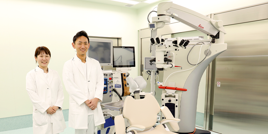
Our dedicated ophthalmology operating room ensures that patients can receive treatment safely and without unnecessary waiting.


Cataracts
Cataracts are a condition that nearly everyone is expected to develop with age. The prevalence is approximately 45% in people in their 50s, 75% in their 60s, 85% in their 70s, and nearly 100% in those over 80 years old.
Our Chief Director has personally performed over 5,000 cataract surgeries to date. He is committed to minimally invasive procedures aimed at enhancing quality of vision (QOV) after surgery. We also offer the latest multifocal intraocular lenses, providing patients with advanced options to reduce dependence on glasses and improve overall visual function.
Retina
In the field of retinal surgery, we are committed to providing ultra-minimally invasive procedures for all types of vitreoretinal diseases. These include retinal detachment, epiretinal membrane, macular hole, intractable proliferative diabetic retinopathy, proliferative vitreoretinopathy, and macular hole retinal detachment associated with pathological myopia. Our approach aims to promote early postoperative visual recovery and minimize complications.
We also perform low-invasive, short-duration surgeries for cases complicated by glaucoma or corneal diseases, utilizing various wide-angle viewing systems and small-incision vitrectomy techniques.
Our Chief Director specializes in vitreoretinal surgery and has performed approximately 2,000 such surgeries—primarily emergency cases—during seven years of service at a university hospital prior to joining our team. He has authored numerous scientific papers, including a doctoral dissertation based on a multicenter study of 27-gauge small-incision vitrectomy for the treatment of primary rhegmatogenous retinal detachment.
Glaucoma
We treat all types of glaucoma, including primary open-angle glaucoma, normal-tension glaucoma, primary angle-closure glaucoma, secondary glaucoma, and congenital glaucoma. While initial treatment typically involves eye drops, surgery may be necessary if intraocular pressure cannot be controlled.
Our services include standard treatments widely adopted across Japan, such as trabeculotomy using a microhook and iStent implantation. We also plan to offer tube shunt surgery and the latest pressure-lowering implant treatments.
Eyelid and Orbit
We provide both medication and incision-based treatments for inflammatory eyelid conditions such as styes (hordeolum) and chalazion. We also perform surgical treatments for common eyelid disorders like age-related ptosis, entropion, exposure keratopathy, and congenital ptosis. For complex eyelid and orbital diseases requiring specialized care, we collaborate closely with oculoplastic specialists at Kyoto Prefectural University of Medicine.
Cornea
We manage a broad range of corneal diseases—from commonly encountered conditions like corneal erosion and herpes simplex keratitis to more challenging ocular surface diseases. We also treat contact lens-related corneal damage and dry eye syndrome. For advanced cases requiring corneal transplantation or other specialized interventions, we work in collaboration with corneal specialists at Kyoto Prefectural University of Medicine.
Main diseases
Cataracts,Epiretinal membrane,Retinal detachment,Diabetic retinopathy,Age-related macular degeneration (AMD),Macular hole,All types of vitreoretinal diseases
Glaucoma,Eyelid disorders,Corneal diseases,Dry eye


Outpatient schedule
Morning 9:00~12:00(Reception8:00-11:30)/
Afternoon to Evening 13:30~16:30(Reception13:00-16:00)
| Mon | Tue | Wed | Thurs | Fri | |
|---|---|---|---|---|---|
| Morning | ● | ● | ● | ● | ● |
| Afternoon to Evening | ● | - | - | - | ● |
Medical track record of the clinical department
-
Surgical method Number of procedures Intraocular Lens (IOL) Implantation / Cataract Surgery 40 Pars Plana Vitrectomy (PPV) – Vitreous Base Dissection under microscope 19 Intraocular Lens (IOL) Implantation / Cataract Surgery 9 Chalazion Excision 1 Pterygium Surgery 1 Removal of Intraocular Foreign Body (Anterior Chamber/Iris) 1 Total number of medical departments 71 -
Anesthesia method Number of cases Local Anesthesia 74 Total number of medical departments 74
Medical Equipment
-
Slit Lamp
Carl Zeiss SL800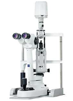
The Zeiss SL800 combines premium optical quality, the renowned Zeiss lens optics, and a high-precision slit illumination system, allowing for high-resolution observation of both the anterior and posterior segments of the eye. The apochromatic optical system significantly reduces chromatic and spherical aberrations, enabling detailed visualization of ocular structures.
-
Ultra-Widefield Scanning Laser Ophthalmoscope
Nikon Silverstone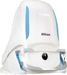
With a 200-degree field of view, this device captures approximately 80% of the retina without pupil dilation and without contact. It can acquire composite color images, red-free images, red-channel images, autofluorescence images, fluorescein angiography images, and indocyanine green angiography images using a single system. It is also equipped with a Swept Source Optical Coherence Tomography (OCT), enabling scans from the posterior pole to the peripheral retina.
-
Optical Coherence Tomography (OCT)
Topcon DRI OCT Triton Pro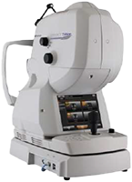
This device provides clear imaging from the vitreous body to the choroid, even in eyes affected by cataracts or vitreous opacities. Equipped with OCT Angiography Ratio Analysis, it enables high-sensitivity and deep penetration imaging of choroidal blood flow, allowing for detailed visualization of vascular abnormalities, including microaneurysms at the capillary level.
-
Optical Biometry Device
Alcon ARGOS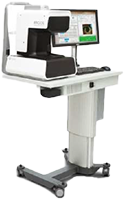
The ARGOS system provides precise preoperative measurements and surgical planning for cataract surgery, including axial length measurement, in a seamless and highly accurate manner.
-
Intraoperative Surgical Guidance System
Alcon VERION
-
Ultrasound Imaging, Biometry, and Pachymetry Device
Tomey UD-800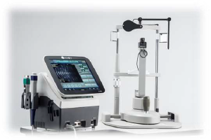
The UD-800 performs ultrasound diagnostic imaging, axial length measurement, and corneal thickness measurement. Its UBM mode enables angle analysis and anterior chamber depth measurement.
-
Skin Electrode ERG
Tomey HE-2000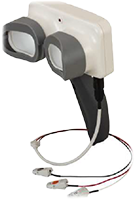
This device allows for ERG measurement using disposable sheet-type skin electrodes, eliminating the risk of corneal infection or trauma. It is particularly useful for patients unable to tolerate corneal electrodes, such as infants, post-surgical patients, and those with corneal disorders. The system enables simultaneous recording of both eyes in a single test.
-
Anterior Segment Optical Coherence Tomography
Tomey CASIA2 Advance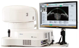
This expert-grade anterior segment OCT supports diagnosis and treatment of cataract surgery, glaucoma management, corneal diseases, and refractive surgery. It provides detailed cross-sectional imaging from the cornea to the lens, including the angle and anterior chamber.
-
Automated Perimeter
Carl Zeiss HFA III 850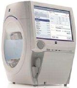
The Humphrey Field Analyzer (HFA) is the gold standard in automated perimetry, widely used in ophthalmology clinics. The HFA III 850 is the latest model, featuring the SITA Faster program, which reduces examination time while maintaining testing accuracy.
-
Multifunction Refractometer
Tomey MR-6000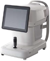
This device integrates five essential examinations in a single unit, including refraction measurement, keratometry, corneal topography, intraocular pressure measurement, and pachymetry.
Additionally, it features a Dry Eye Analysis App, which assists in diagnosing dry eye syndrome, a condition affecting approximately 22 million people due to aging, air conditioning, prolonged screen use, and contact lens wear. -
Auto Lens Analyzer
Topcon SOLOS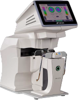
The SOLOS auto lens analyzer enables fully automatic, simultaneous measurement of both eyes, providing detailed data on lens type, power, axis, and transmittance. It also visualizes progressive and multifocal lens power maps for enhanced analysis.
-
Pattern Laser
Topcon PASCAL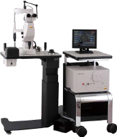
Developed in collaboration with Stanford University, PASCAL features a unique laser technology that performs retinal photocoagulation in a shorter duration with higher energy output, minimizing damage to the inner retina and choroid while reducing patient discomfort.
The pattern laser technology shortens the treatment time for panretinal photocoagulation (PRP), reducing the burden on both surgeons and patients. -
Tango Ophthalmic Laser
Ellex Tango PRO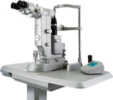
This high-performance laser system is designed for glaucoma and cataract treatments. It features a newly designed laser head with superior durability and stable output, enabling precise and reproducible treatments.
- The Selective Laser Trabeculoplasty (SLT) technology by Ellex ensures high-precision energy control, providing extremely accurate SLT procedures.
- The YAG mode enables capsulotomy and iridotomy with efficient, low-energy pulses, reducing the risk of lens pitting, retinal thickening, intraocular pressure spikes, and collateral tissue damage.
-
Ophthalmic Surgical Microscope
EnFocus Intraoperative OCT Imaging System & Leica Proveo 8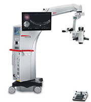
The Proveo 8 offers advanced ophthalmic microscopy solutions, supporting anterior and posterior segment surgeries for better patient outcomes. The EnFocus OCT system provides real-time intraoperative imaging, enabling surgeons to assess fine structural changes and optimize surgical plans accordingly.
-
Cataract Surgery System
Alcon CENTURION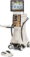
Designed for challenging cataract cases, the CENTURION system maintains near-normal intraocular pressure (IOP) and high anterior chamber stability, enhancing intraoperative safety and reliability. By combining Ozil technology and advanced fluidics, it ensures efficient and gentle cataract removal, providing a safer surgical experience for patients.
-
Vitrectomy System
DORC EVA NEXUS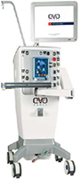
The EVA NEXUS vitrectomy system synchronizes the surgeon, system, and patient conditions using advanced fluidic control and VTi (VacuFlow Venturi) technology.
- The SMART IOP feature automatically detects and compensates for pressure fluctuations, maintaining stable anterior chamber conditions even after occlusion breakage.
- The VTi pump eliminates unnecessary pulsation risks, ensuring consistent intraocular pressure (IOP) stability during vitreoretinal surgeries with its next-generation active infusion system.
Regarding clinical research
Click here for more information on clinical research